Start date: Week 39 (24th September 2024)
Speaker: Daniel Fürth (Assistant Professor, SciLifeLab Fellow; Tuesday 24th September 8:00-10:00 CEST)

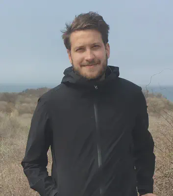
Dr. Daniel Fürth completed his PhD at the Department of Neuroscience, Karolinska Institute, Sweden, where he developed computational methods for 3D tissue reconstruction and connectomics. During his postdoc at Cold Spring Harbor Laboratory (CSHL) in New York, USA, he developed single-molecule RNA imaging techniques. Now, as a group leader, SciLifeLab fellow, and assistant professor at the Department of Immunology, Genetics & Pathology, Uppsala University, Dr. Fürth and his team develop novel sequencing chemistries and imaging approaches to detect molecules directly in living cells.
Title: Single-Molecule Analysis and Tissue Atlases
This lecture will explore advanced techniques in image segmentation and registration, focusing on their application in creating comprehensive tissue atlases. We will begin by discussing standard segmentation methods, including the use of deep learning architectures such as ResNet and U-Net, which are widely employed in medical imaging for precise delineation of anatomical structures. We will then move to registration techniques, with a particular focus on point-set registration, which aligns multi-dimensional datasets to build accurate reference atlases. Best practices for constructing reference atlases will be covered, including the integration of ontologies for labeling anatomical regions, enabling the conversion of 2D histological data into 3D tissue maps. Lastly, we will examine tracking methodologies for single-molecule and cell dynamics over time, highlighting techniques that allow for detailed spatial and temporal analysis of biological processes. This comprehensive overview will provide attendees with key insights into the development of tissue atlases and the tools needed for tracking and analyzing molecular behavior across time and space.
Computer Lab: The lab session will involve hands-on exercises using Jupyter notebooks to apply the discussed computational tools for analyzing fluorescent microscopy data. Participants will practice segmenting and tracking single molecules, as well as performing tissue atlas registration.